If we want to understand how the recovery after Bell’s palsy happens, we must thoroughly know the structure of the facial nerve first. Learning about the structure and function of the facial nerve will help you to understand what is a conduction block (explained below). Only then we can analyze and choose proper rehabilitation methods that are able to provide the desired results.
The function of the facial nerve
Our facial nerve can be easily compared to a thick telephone cable that connects the telephone station (nerve nucleus in the brain) with the neighbourhood of the town (our face). Large bundles of wires split to reach separate apartment blocks (parts of the face). Then they go to various floors of the apartment blocks (facial muscles). Finally, the individual wires supply phone signals to each separate apartment (muscle fibres, lacrimal gland and taste sensors).
The primary job of the facial nerve is to supply electrical signals from the brain to the respective muscles fibres in our facial muscles. When those signals reach via the nerve to the fibres, they contract or relax, producing our facial expressions.
Although our facial nerve is less than 1 millimetre thick, it contains 10,000 nerve cells (neurons).
There are 3 sorts of neurons in the facial nerve.
Sensory neurons are responsible for the taste receptors in a part of our tongue. That is why when Bell’s palsy happens, some patients have a weird taste sensation or a loss of taste on one side of the tongue (anterior two thirds).
Parasympathetic fibres are responsible for the function of the tear gland. During the acute phase of Bell’s palsy, our affected eye becomes dry because the tear production signals cannot reach the lacrimal gland.
Motor neurons are very important. They connect our brain to the facial muscles, so we can blink, reflect our emotions and produce articulated speech. Motor neurons are combined in bundles that supply mimetic signals to various parts of the face. Those bundles are divided into smaller ones to manage individual facial muscles, and then further divided into very small bunches that regulate the function of parts of muscles. Individual neurons (nerve fibres) supply mimetic signals to single muscle strands, just like phone calls that arrive to your home or to a mobile phone. There are about 7,000 motor neurons in the facial nerve.
The structure of the facial nerve (motor neuron)
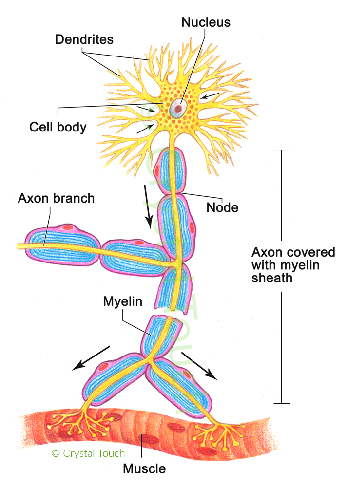
A motor neuron consists of 3 important parts:
- cell body (soma),
- dendrites (numerous branch-like connections to exchange signals between individual neurons),
- and the axon – a long “wire” that connects the cell body with the corresponding muscle fibre.
The Axon
Axons are the highways between the brain and the facial muscles. They conduct finely coordinated signals from the facial nerve nucleus to various groups of muscle fibres to produce our facial expressions.
The axon ends at the neuromuscular junction. It is the very narrow gap between the nerve and muscle tissue, where the electric signal passes on from the nerve fibre to the muscle fibre and excites it. This excitation results in contraction.
What is a conduction block?
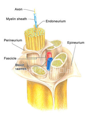
Just like any electric wire, each individual nerve fibre (axon) is electrically insulated with the sheath made of a special protein called myelin. It is also mechanically protected by a tube-like endoneurium. When Bell’s palsy happens, myelin gets damaged firstly from a mechanical compression by the swollen tissue, electrical insulation is lost, and mimetic signals cannot reach the facial muscles. This is called a conduction block.

Conduction block can also be a result of the effect of toxins. For example, Botulinum Toxin (Botox) permanently blocks neuromuscular junctions. This damage is irreversible. So to recover its function, the affected axon needs to grow side-terminals to regain the connection with the corresponding muscle fibres. This explains why the effect of Botox injections is only temporary. Depending on the regenerative capacity of the body, within 2 to 5 months facial nerve branches re-connect to “their” facial muscles and restore their functions.
It is worth mentioning that during the whole period after Botox injection, mimetic signals from the brain are still produced. They go all the way along the axons to the muscles, but cannot pass the blocked neuromuscular junctions.
At Crystal Touch we work on “fixing” the signals originating in the brain, to make sure the right mimetic signals arrive at the muscles, leading to long-lasting results.