Crystal Touch Bell’s palsy clinic often participates in various congresses and conferences for neurology and neuro-rehabilitation. Our purpose is to shift the paradigm for rehabilitation of residuals and complications of long-standing facial palsy in general, and of synkinesis in particular.
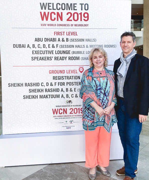
The World Congress of Neurology 2019 took place in Dubai. We were delighted to present our findings there, as well as to discuss and share our ideas with other neurologists.
The topic of our presentation at WCN 2019 was:
“Facial synkinesis in patients with long-standing Bell’s palsy is a reversible consequence of disrupted reciprocal inhibition caused by prolonged absence of proprioceptive feedback”.
Our presentation at WCN 2019
We suggested a new underlaying cause of synkinesis, different from traditional and currently prevailing theory. Watch the video below to see our presentation, or read the transcript and explanations under the video to learn more about our approach to rehabilitation of facial synkinesis after a long-standing facial palsy.


New paradigm for rehabilitation of synkinesis
Synkinesis, is by far the most devastating complication of long-standing Bell’s palsy. It has a dramatic effect on a patient’s quality of life, may lead to a loss of self-esteem, loss of self-confidence, sometimes result in social isolation and can even result in suicidal behaviour.
Traditional paradigm in the rehabilitation of facial palsy suggests that if after one or two years there was no full recovery, then further improvements are not to be expected. The patients often hear from their physician that nothing can be done any more, and that they must learn to live with the residuals and complications.
We think differently about this issue.
The considerations that we are sharing with you, may suggest that if the new paradigm receives support and confirmation from subsequent research, it can lead to substantial changes in the rehabilitation of the patients with residuals and complications of facial palsy.
The traditional paradigm
In 1975, Japanese professor Jun Kimura, now working at Iowa State University, published a fundamental paper “Electrophysiologic analysis of aberrant regeneration after facial nerve paralysis”. Since then, the aberrant regeneration is considered by most specialists to be the main cause of facial synkinesis.
The traditional paradigm leaves both the patient and the specialist with a limited choice of methods and techniques that are aimed mainly at the management of symptoms and do not address the fundamental causes of synkinesis.
The main notion of aberrant regeneration is that during the recovery process, fibres of the facial nerve regrow and reconnect to the wrong facial muscles. That’s why when we’re blinking or smiling, other muscles become involved, and we cannot produce the desired mimetic expression.
Our consideration regarding the cause of this inability to produce desired mimetic expression is very different.
The new paradigm considerations
At Crystal Touch clinic, we continue to research synkinesis after facial palsy. Based on the experience of our patients, we see that it is possible to improve and reduce synkinesis. If aberrant regeneration were the true cause of synkinesis, and the nerve fibres would grow to the wrong facial muscles, such improvements would not be possible.
Below are a few reasons and discussions for the new paradigm for the origins of synkinesis.
Quantitative measurement of synkinesis

At Crystal Touch, we developed an instrumental test for Synkinetic Correlation. It shows that the efforts of facial muscles on the affected side are 2 to 5 times greater than those on the healthy side for the same facial movements.
On the image, you can see two sides of the face of one of our patients. On the left-hand side – the healthy side – you can see that when she closes or blinks her eyes, this side does not show a lot of effort. The affected side, on the right-hand side of the image, is very active in both muscles around the eye and around the mouth corner. It is even more visible when the patient makes a rhythmical broad smile. Then the difference between the healthy and the affected side is even greater.
Volitional and emotional control of mimetic signals
At any given moment, our face reflects an overlap of two mimetic spectra: one coming from the emotional centre of the brain and the other – from volitional one.
Emotional signals happen automatically, as an emotional response. The volitional signals are what we consciously create.
When we experience an emotion, our brain forms mimetic signals to produce the corresponding facial expression. Proprioceptive signals from facial muscles, skin and facial tissues serve as sensory feedback. These signals travel back to our brain, where it compares this received information with the ideal pattern that is stored in its “library” of emotions. The brain then will take incremental corrections to the contraction signals until the feedback from the face matches the ideal pattern of emotion in the “library” and we express the desired emotion with respective facial movements.
After a long-standing facial palsy, the facial movements carry a very large volitional component. This means the patient’s brain uses mimetic amplification all the time for whichever kind of facial movements it is engaged in.
Muscles-antagonists
While analysing the pictures of 13 standard facial expressions of more than 800 of our facial palsy patients, we have noticed that along with synkinesis, we can often observe simultaneous contractions of antagonist muscles.
In this example (image with Example 1), we can see that when the patient is squeezing her eyes tightly, then on the affected side her m.frontalis contracts involuntarily, overpowers the m.orbicularis oculi and pulls up the eyebrow instead of squeezing the eye. From our point of view, this is a result of motor overflow and lack of reciprocal inhibition on the affected side.

The same happens during the broad smile expression (image with Example 2). When the patient smiles, together with the zygomatic muscles, their antagonist-muscle which is m. Depressor Anguli Oris, and also m.mentalis contract involuntarily, pressing down the mouth corner and, thus, making the smile asymmetrical.
Simultaneous contraction of antagonists during facial movements, from our point of view, again, is a result of a disruption in reciprocal inhibition.
Synkinesis as a “side effect” of motor overflow

Practically all patients after long-standing facial palsy develop a certain type and a certain degree of facial synkinesis. From our point of view, synkinesis can be regarded as a kind of “side effect” of disrupted reciprocal inhibition of muscles-antagonists.
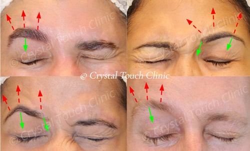
On the image below, you can see the vectors (blue arrows) of the contraction direction of facial muscles.


This demonstration shows how the co-contraction of antagonists (red arrows) distorts the facial expression on the affected side.
Motor overflow from the maximal effort results in synkinetic contractions of “muscles-followers”. In this case, it is the Zygomatic muscles. When we are squeezing our eyes tightly, then m. Orbicularis Oculi becomes the muscle-“driver”, and m. Zygomatici are the “followers” in this synkinetic movement.
During the long recovery, due to lack of sensory feedback, a constant volitional amplification of facial muscles contractions becomes a “habit” of the brain.
When the regenerating axons reach facial muscles, then the proprioceptive feedback and repetition gradually consolidate synkinetic facial pattern into a conditioned reflex.
Our suggested new paradigm
The suggested new paradigm for the rehabilitation of synkinesis is from our point this:
- Synkinesis is a pathological mimetic pattern that is formed in the volitional mimetic centre of the brain.
- Synkinesis is a “side effect” of disrupted reciprocal inhibition caused by continuous lack of proprioceptive feedback from the face, and by constant cortical amplification of facial muscles.
- Synkinesis is essentially a conditioned reflex.
- As any conditioned reflex, synkinesis is per definition reversible by prolonged negative feedback.
We should welcome the new methods, new techniques and new approaches to rehabilitation of long-standing facial palsy. We should welcome the emergence of new, substantiated hope for the millions of facial palsy patients to improve their facial symmetry, to bring back their long-lost smiles and the most important – to substantially improve their quality of life.